
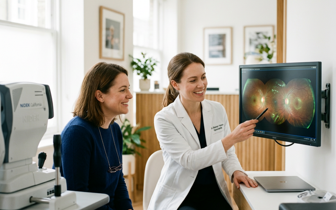
Medically reviewed by Dr. Alisha Seger, OD • Updated November 2026
When you sit down for an annual eye exam, you might expect a standard vision check to determine if your prescription has changed. However, modern optometric diagnostic equipment does far more than evaluate visual acuity. It provides an unobstructed, high-resolution view directly into your body’s vascular and neurological systems.
At The Spectacle Shoppe in Franklin, TN, we utilize Optomap ultra-widefield digital retinal imaging. In less than a second per eye, this technology captures a high-definition, 200-degree panoramic view of your retina. This covers approximately 82% of the retinal surface in a single sweep, compared to the 30 to 45 degrees captured by traditional desktop cameras.
Because the retina is the only place in the human body where live blood vessels and nerve tissue can be observed directly without invasive surgery, a digital retina scan frequently detects systemic health conditions long before symptoms appear elsewhere in the body.
1. High Blood Pressure (Hypertension) & Hypertensive Retinopathy
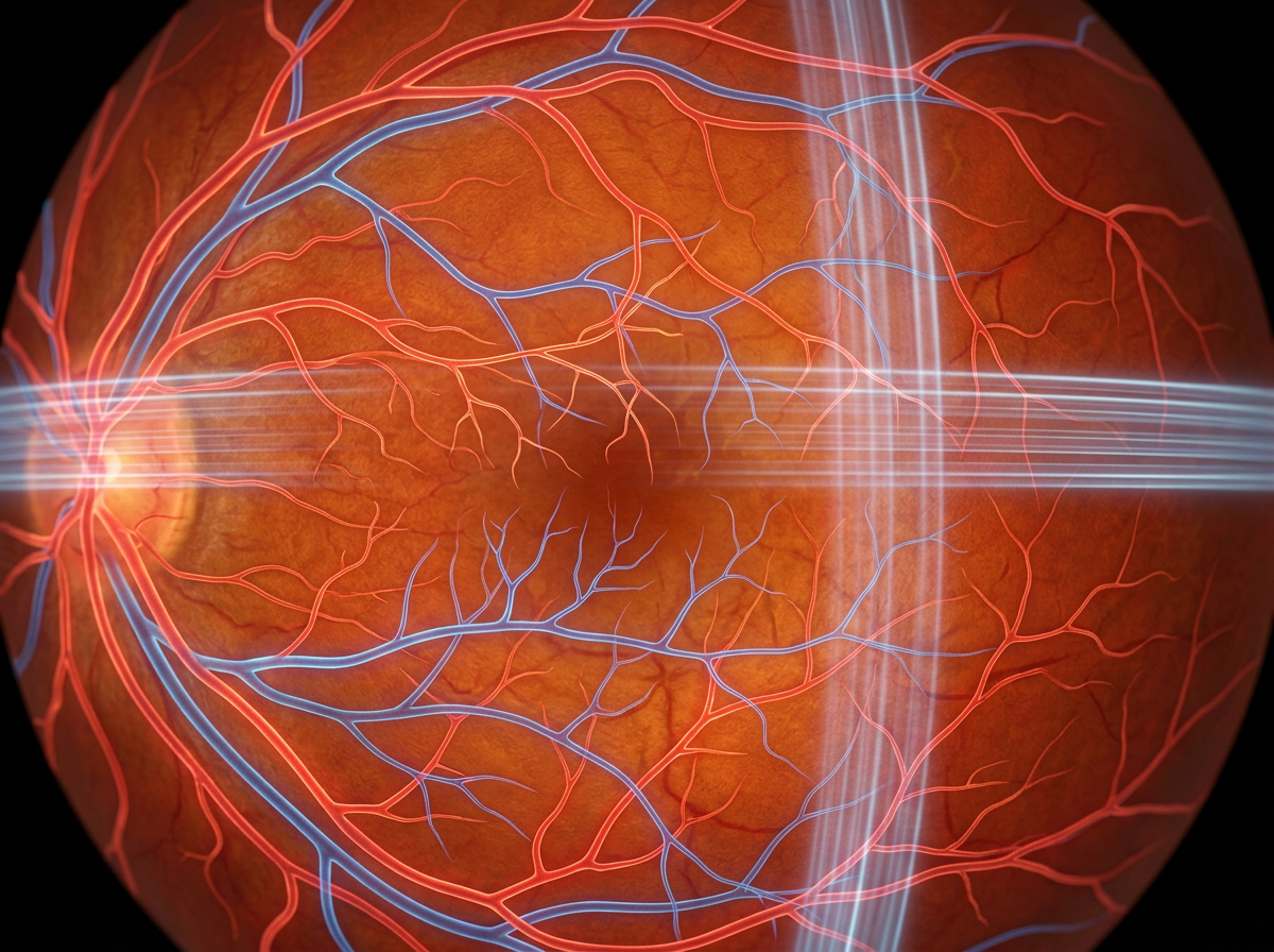
Your retinal blood vessels mirror the vascular health of your heart, brain, and kidneys. High blood pressure damages these delicate microvessels over time, leading to structural changes that are visible during an Optomap scan.
What the Scan Detects:
- Arteriolar Narrowing & “Copper or Silver Wiring”: Chronic elevated pressure causes arterial walls to stiffen, thicken, and reflect light differently under digital imaging, giving them a metallic appearance.
- Arteriovenous (AV) Nicking: Stiffened arteries cross over retinal veins and compress them, restricting return blood flow.
- Flame-Shaped Microhemorrhages: High blood pressure can cause tiny capillary walls in the nerve fiber layer to rupture, leaking small amounts of blood onto the retina.
Clinical Significance: Because hypertension is largely asymptomatic in its early stages, an Optomap scan often serves as the initial trigger for a patient to seek cardiovascular evaluation from their primary care physician.
2. Diabetes & Diabetic Retinopathy
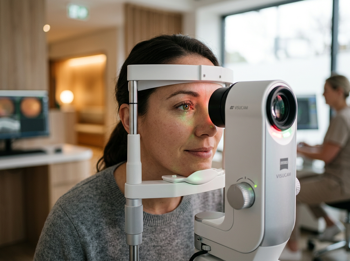
Diabetic retinopathy is a leading cause of preventable blindness in American adults. High blood sugar damages the microvascular endothelial cells lining the small blood vessels of the retina, causing them to leak or become blocked entirely.
What the Scan Detects:
- Microaneurysms: Small, balloon-like bulges in the capillary walls that appear as distinct red dots on high-resolution digital scans.
- Hard Exudates: Yellowish deposits of lipid and protein leakage that escape from damaged, hyperpermeable blood vessels into the deep retinal layers.
- Cotton Wool Spots: Pale, white patches indicating areas where microvascular blockage has deprived the retinal nerve fiber layer of oxygen (retinal ischemia).
- Neovascularization: Advanced diabetic retinopathy triggers the growth of fragile, abnormal new blood vessels that bleed easily into the vitreous gel.
Clinical Significance: Ultra-widefield imaging allows our optometrists to monitor subtle changes across the peripheral retina, enabling timely intervention before central vision is compromised or permanent diabetic macular edema develops.
3. Glaucoma: The Silent Thief of Sight
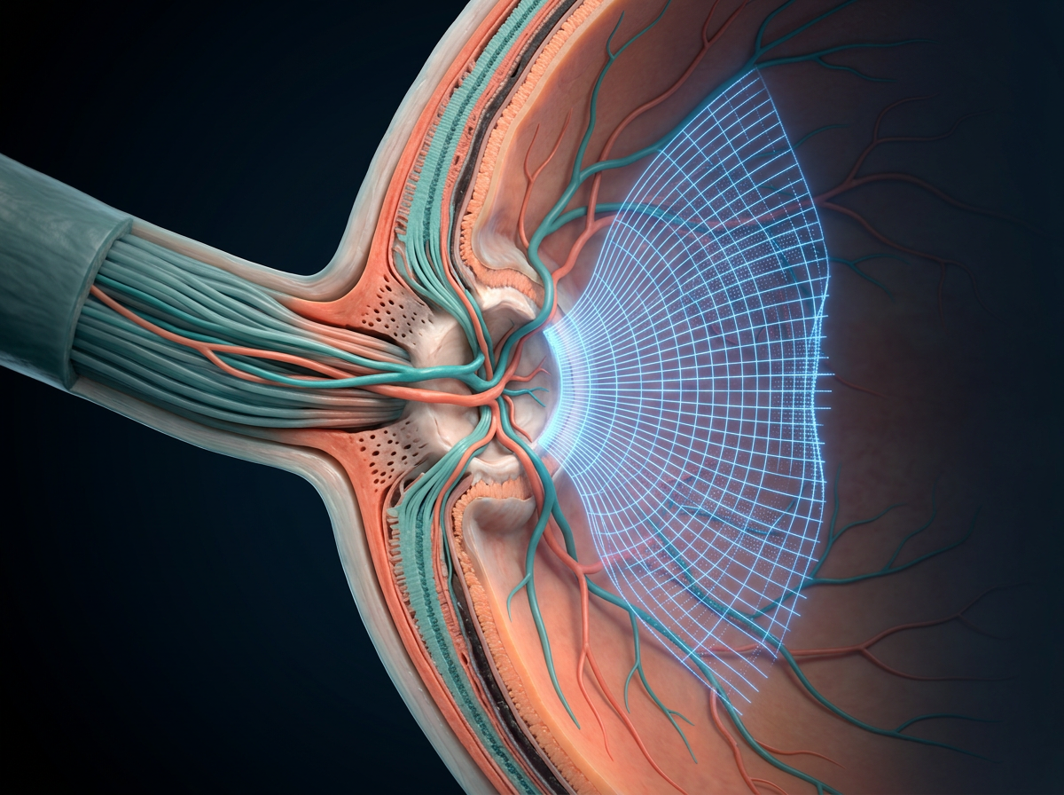
Glaucoma is a group of eye conditions that damage the optic nerve, often driven by elevated intraocular pressure (IOP). Because peripheral vision loss occurs gradually, often starting in the far edges of your sight field, most patients notice no symptoms until irreversible damage has occurred.
What the Scan Detects:
- Optic Nerve Head Cupping: The Optomap scan captures detailed imaging of the optic disc, allowing doctors to evaluate the cup-to-disc ratio. As nerve fibers die off from pressure, the central cup within the optic nerve head enlarges.
- Neuroretinal Rim Thinning: Digital imaging tracks localized thinning or notching of the healthy neuroretinal tissue encircling the optic disc.
- Retinal Nerve Fiber Layer (RNFL) Defects: Digital filters reveal subtle wedge-shaped dark spots where bundles of nerve fibers have degenerated.
Clinical Significance: Baseline digital imaging creates a precise digital baseline. By comparing annual Optomap images side-by-side over time, doctors can detect subtle microscopic changes long before standard field-of-vision tests reveal noticeable loss.
What to Expect During Your Optomap Scan
| Feature | Details |
| Duration | Under 1 second per eye capture time (less than 3 minutes total setup). |
| Dilation | In most patients, no dilating eye drops are required. |
| Comfort | Completely non-contact and pain-free, using a low-intensity flash of red and green laser light. |
| Review | Results are rendered immediately on screen for review during your appointment. |
Schedule Your Annual Comprehensive Exam in Franklin
Your eye health provides a vital window into your total body wellness. To schedule your annual eye exam with Dr. Alisha Seger or Dr. Jason Chu at The Spectacle Shoppe, visit our Franklin clinic location at 1624 W Main Street or call 615-599-4460.
Recent Comments